Dental Imaging and Diagnostic Equipment
Written by the Equate Instruments Technical Team · Reviewed by the Medical & Dental Board, Equate Instruments · Updated ·
Dental imaging and diagnostic equipment is everything used to make, process and read radiographs of the teeth and jaws: the X-ray unit, film or digital receptors, bitewing tabs and film holders that position them, shielding and staff dosimeters, darkroom or automatic processing gear, view boxes, panoramic, cephalometric and cone-beam CT machines, and adjunct aids for examining the oral mucosa.
Key points
- Every radiograph needs three things lined up: a source (the X-ray tube head), a receptor (film, a digital sensor or a phosphor plate) and a holder that keeps the receptor still and correctly angled.
- European, UK and US guidance now puts justification, rectangular collimation and fast receptors ahead of patient shielding. Routine aprons are no longer recommended, although UK guidance keeps a thyroid shield where the gland is unavoidably in the primary beam, and IAEA guidance where it is in or near the main beam; national regulations decide what a clinic must do.
- Receptor holders are heat-sterilised between patients, while sensors and phosphor plates are covered with a single-use barrier and cleaned as their manufacturer specifies.
What is dental imaging and diagnostic equipment?
Dental imaging covers far more than the X-ray machine on the wall. Linda Bartolomucci Boyd's Dental Instruments: A Pocket Guide (Elsevier) gives this group a chapter of its own, running from intraoral film and the tabs and holders that position it, through shielding, processing and viewing, to digital sensors, panoramic and cephalometric units, cone-beam CT and a light-based aid for checking the oral mucosa.
The kit sorts into four jobs: making the exposure, capturing the image, turning it into something readable, and keeping staff, patients and equipment safe. Radiographs add to the visual and tactile examination with mirror and explorer described in the basic dental instruments guide; they don't replace it, and radiograph holders join the basic dental tray only when images are planned. The table below groups the main items by job, and the sections that follow take each family in turn.
| Job | Equipment | Notes |
|---|---|---|
| Making the exposure | Intraoral X-ray unit (control panel, tube head, round or rectangular PID), handheld portable unit | Exposure is set by tooth region and patient size; a rectangular PID narrows the beam |
| Capturing the image | Intraoral film (sizes 0–4), wired or wireless digital sensors, phosphor storage plates | Digital receptors need no chemistry and generally cut dose against film, but some phosphor plates are no faster than D-speed film, and a plate's wide latitude can hide an overexposure |
| Positioning the receptor | Bitewing tabs, bite-block holders, ring-and-arm paralleling holders | Holders improve geometry and stop staff holding film in the mouth |
| Processing and reading | Manual tanks, film racks and hangers, safelight, automatic processor, view box, duplicator, phosphor plate scanner, imaging software | Film needs a darkroom or daylight loader; digital images appear on screen |
| Protection | Collimation, staff dosimeters, aprons and thyroid collars where required, barriers | Requirements are set by national radiation and infection-control rules |
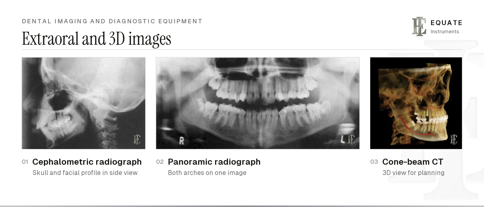
| Extraoral and 3D | Panoramic, cephalometric and cone-beam CT units | Used for whole-arch, skeletal and three-dimensional assessment |
Intraoral film and receptor sizes
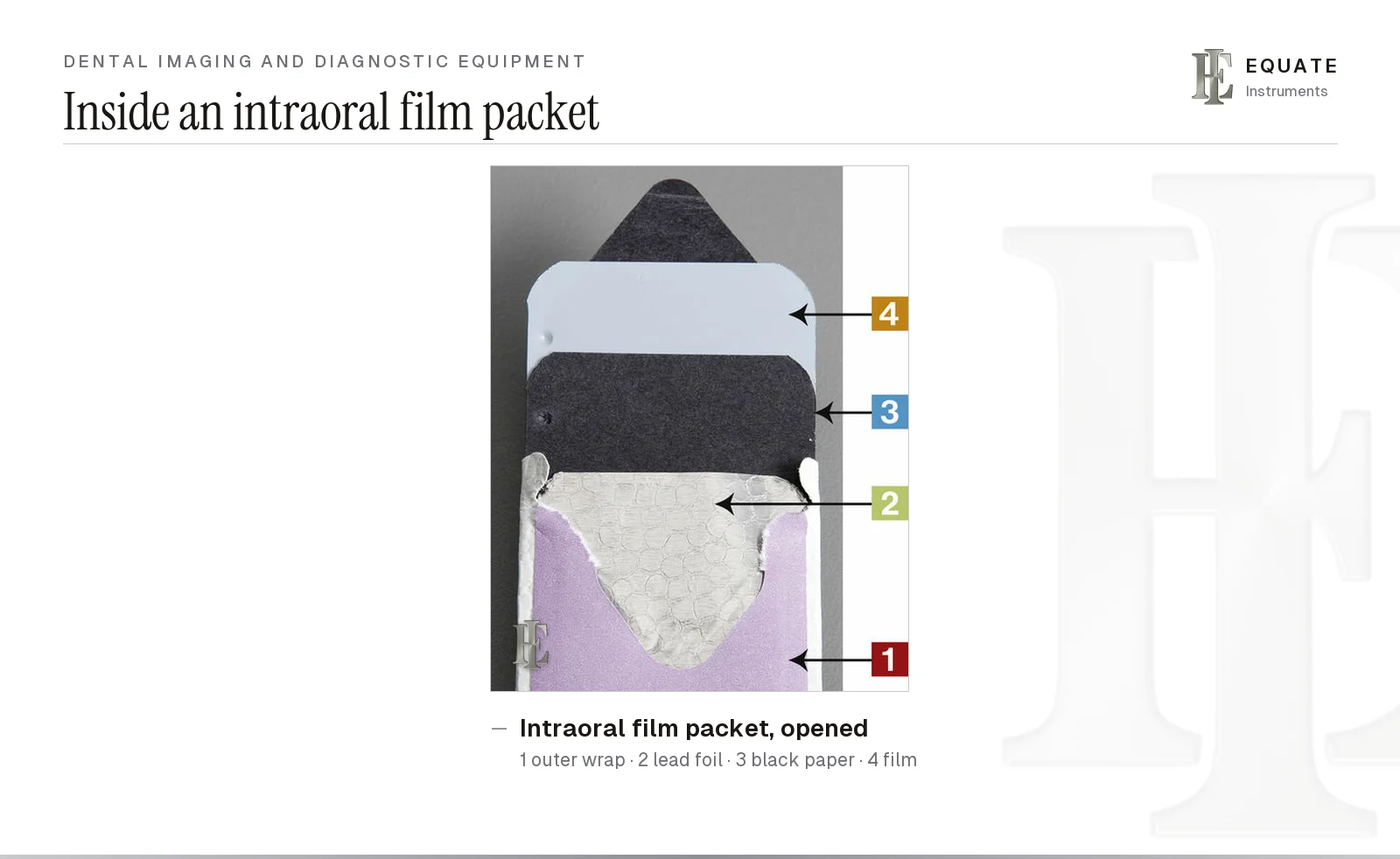
Conventional intraoral film comes sealed in a light-tight, moisture-resistant packet. Inside, the film itself (single or paired, so that a duplicate is made at the same exposure) is wrapped in black paper and backed by a thin sheet of lead foil, which absorbs radiation that has passed through the film and reduces fogging from scatter. The plain side of the packet faces the teeth and the tube; the side with the opening tab faces the tongue or palate.
Each film carries a small embossed dot. Its raised (convex) side faces the tube, which is how a processed film is later mounted the right way round, and the dot is placed towards the biting edge so it doesn't fall over a root apex. Film speed is graded by letter, and the faster the film, the lower the exposure it needs; D, E and F are the intraoral grades. UK guidance from Public Health England and FGDP(UK) (2020) says D-speed film gives high patient doses and should no longer be used, and that a clinic still using film should choose F speed, which is roughly 20% faster than E. European guidance in Radiation Protection 136 likewise advises the fastest film available or a digital receptor.
Every box states the film type and speed, how many films each packet holds and an expiry date. Rotate stock so the oldest is used first, and keep it somewhere cool and dry, well away from the X-ray unit: heat, humidity, age and stray radiation all fog film before it is ever exposed.
| Size | Typical use | Comment |
|---|---|---|
| 0 | Periapicals and bitewings in young children (roughly under 3) | Smallest packet; easier for a small mouth to tolerate |
| 1 | Anterior periapicals in adults | Narrow shape fits the curve of the front of the arch |
| 2 | Adult posterior periapicals and bitewings; anterior periapicals too | The workhorse size in most practices |
| 3 | Extended (long) bitewings | Rarely stocked now |
| 4 | Occlusal views of the upper or lower arch | Largest intraoral film; the patient bites gently on it |
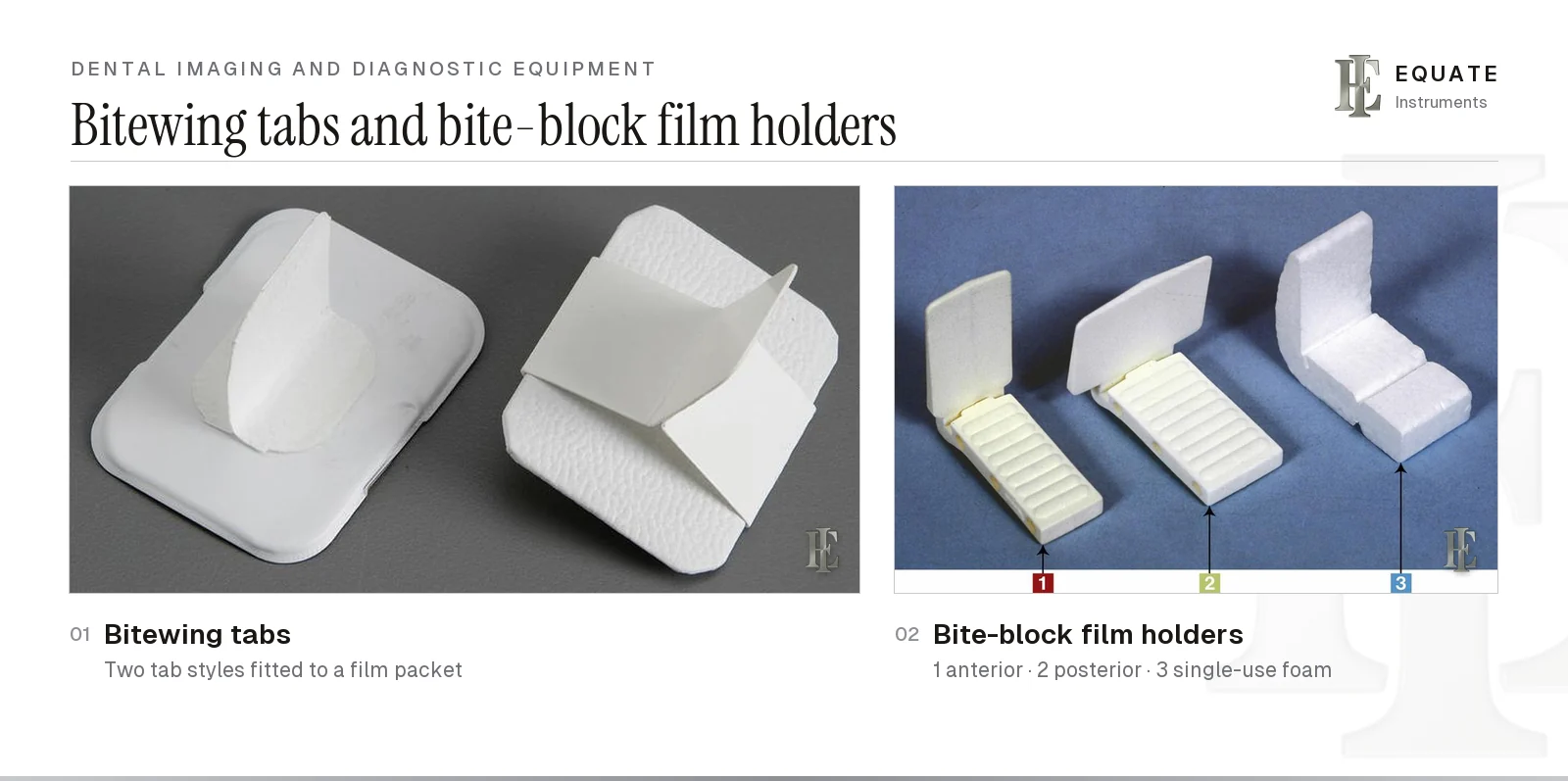
Bitewing tabs and periapical film holders
A bitewing shows the crowns of upper and lower back teeth on one image and is mainly used to find caries between teeth and to check bone levels and restoration margins. The simplest way to take one is with a tab: a card or plastic wing that either sticks onto the packet or slides over it. The patient closes on the wing to hold the film against the teeth. Tabs are single-use and are discarded after the exposure. Depending on the patient and local selection criteria, a set is usually two or four bitewings, covering the premolar and molar regions on each side.
Periapical radiographs show the whole tooth, from crown to apex, with the surrounding bone. Simple bite-block holders have a slot that grips the film and a ridged block for the patient to bite on; reusable plastic versions are made in anterior and posterior shapes, and foam blocks are made for single use. Double-ended plastic holders put an anterior and a posterior slot on the same piece. They are quick to use but give no guide to tube alignment, so the operator lines up the PID by eye.
Whatever holder is used, the receptor should not be held in place by a member of staff. If a patient genuinely cannot hold a receptor, national rules set out who may help and what protection they wear.
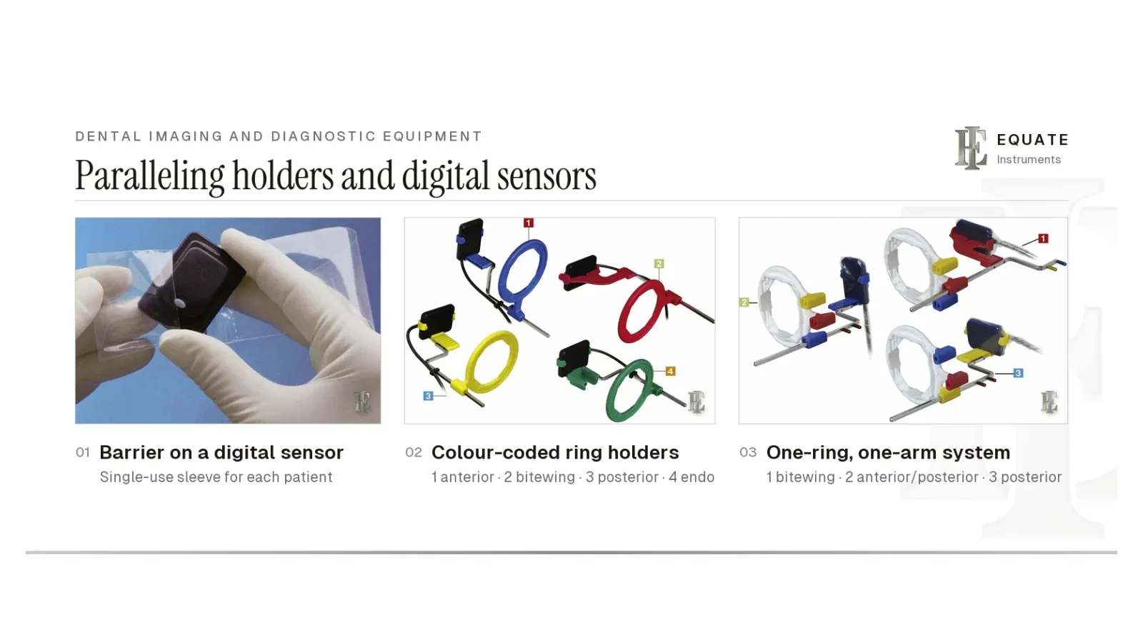
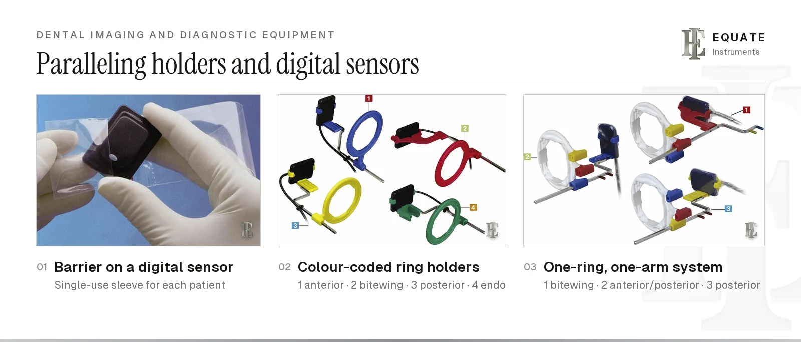
Paralleling holders and digital sensors
Ring-and-arm holders are built for the paralleling technique: the receptor is placed parallel to the long axis of the tooth and the X-ray beam is aimed at right angles to both. A bite block holds the film or sensor, a metal indicator rod runs out of the mouth, and a sighting ring slides along the rod. When the end of the PID is lined up with the ring, the beam is correctly angled and centred, which reduces cone cuts, foreshortening and elongation. The older alternative, the bisecting-angle technique, rests the receptor against the crown and aims the beam at right angles to an imaginary line halving the angle between tooth and receptor. It helps where a shallow palate or small mouth prevents parallel placement, but it distorts root length more easily. UK guidance asks for paralleling on periapicals unless there is a clear clinical reason not to, because it gives better, more repeatable images at a lower dose.
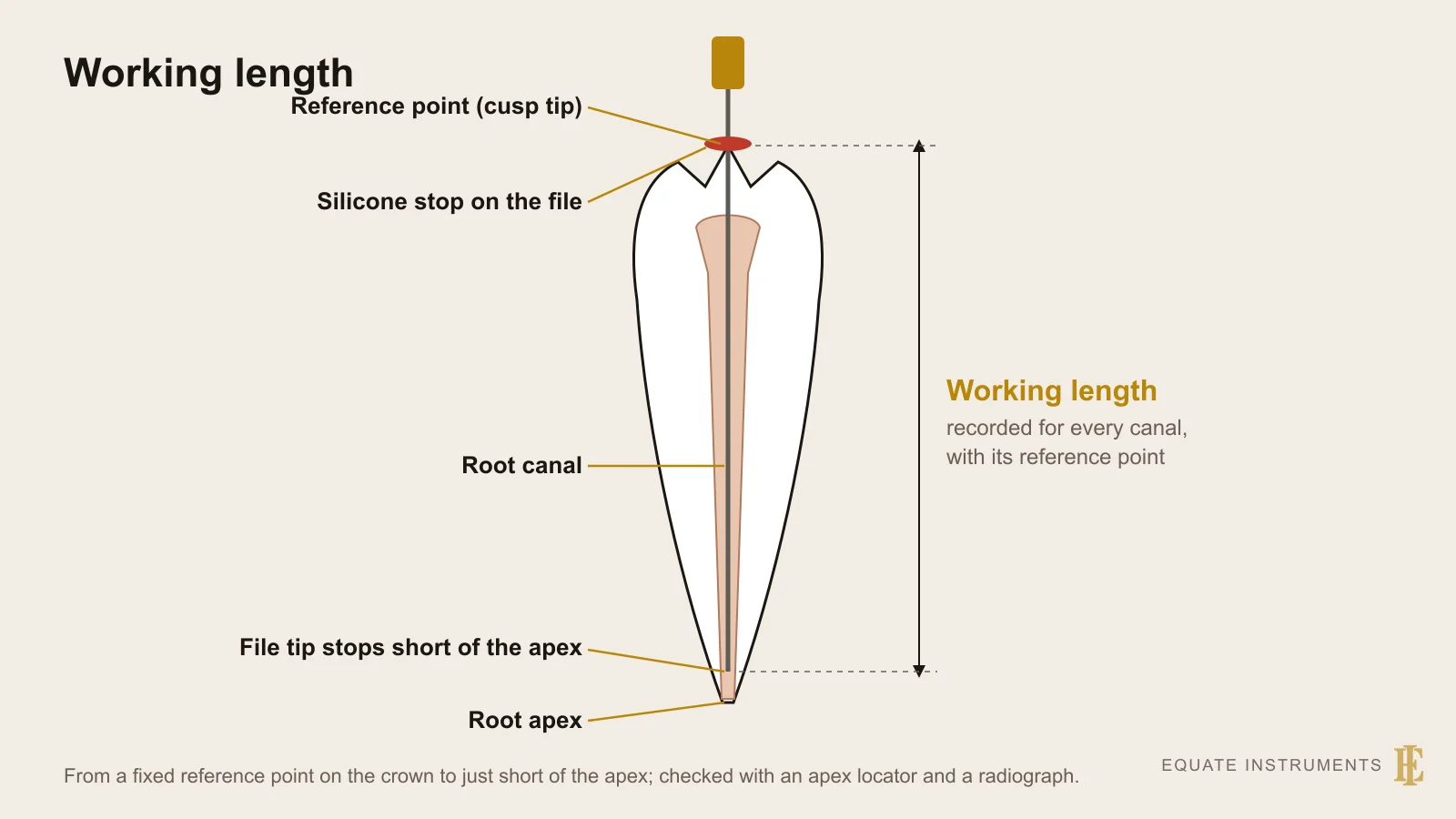
Most systems follow the same colour code: blue for anterior periapicals, yellow for posterior periapicals, red for bitewings and green for endodontic views, where the holder leaves room for files and clamps and a radiolucent rubber dam frame can stay in place for the exposure. One-ring systems carry a single ring and arm with colour-coded slots, so fewer parts need to be stocked and reprocessed. Holders for digital sensors look the same but have deeper, grippier slots and room for the cable. An apex locator now sets working length in many cases, and UK selection criteria call for a radiograph with a file in the canal when that reading is in doubt; for the steps, see the endodontic instruments guide and the endodontic tray setup. The length is then carried to each file with a dental measuring ruler.
Digital receptors replace film in most clinics. Direct sensors are rigid, wired or wireless electronic plates that send the image to the computer within seconds. Phosphor storage plates are thin and flexible like film; after exposure they are read in a scanner, which then erases them for reuse. They need no darkroom, though an exposed plate should be kept out of bright light until it is scanned. Both are covered with a fresh barrier sleeve for each patient. Sensors come in a few sizes that roughly match common film sizes, and their thickness and rigid edges can make placement less comfortable than film.
The intraoral X-ray unit and radiation protection
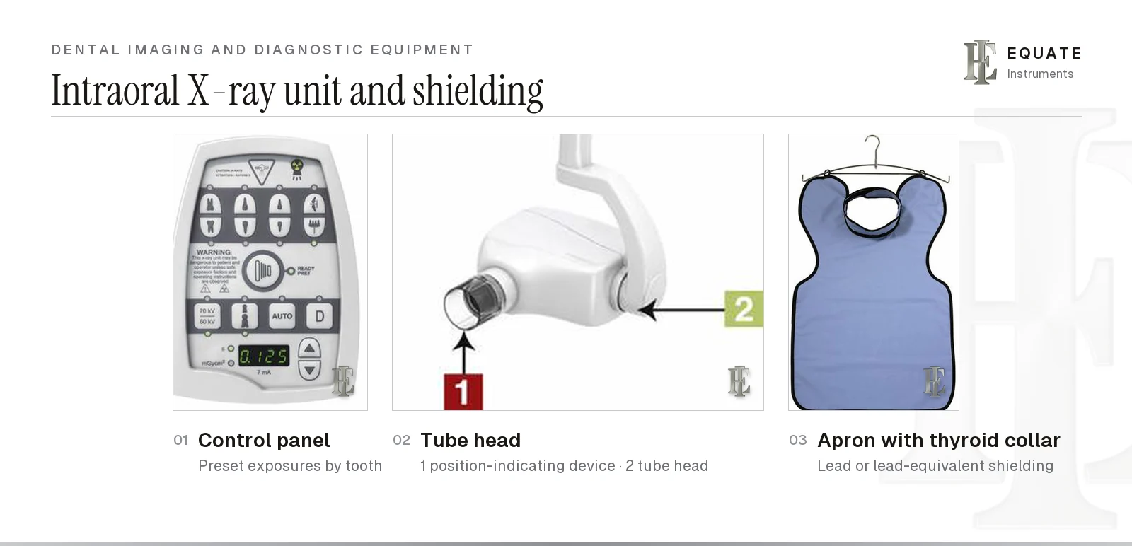
A wall- or chair-mounted intraoral unit has three main parts: the control panel, where exposure is selected; the tube head, which produces the beam; and the position-indicating device (PID), the open-ended cylinder or rectangle that points the beam at the receptor. Many panels have preset exposures by tooth region and patient size; others also allow kilovoltage (kV) and tube current (mA) to be set. The exposure switch should sit where the operator can stand outside the controlled area, or behind a barrier, during the exposure. UK guidance prefers designating the whole room; where that is not practicable, the controlled area is everything within 1.5 metres of the tube and the patient while the set is ready to fire, plus the path of the primary beam. For CBCT, only whole-room designation is recommended.
Rectangular PIDs collimate the beam close to the size of the receptor and cut the area of tissue exposed. The European Commission's Radiation Protection 136 and the American Dental Association's 2024 recommendations both favour rectangular collimation, fast or digital receptors and, above all, justification: an image is taken only when it is expected to change diagnosis or treatment. The UK Guidance Notes for Dental Practitioners (Public Health England and FGDP(UK), 2020, written under IRR17 and IR(ME)R17) put numbers on it: a rectangular collimator roughly halves effective dose compared with a round cone, so it should be used for periapicals, bitewings and occlusals unless a documented clinical reason prevents it. For a size 2 receptor the beam at the cone tip should measure no more than 40 × 50 mm, ideally under 35 × 45 mm. The same guidance expects routine equipment tests at intervals of no more than three years. Handheld portable units with internal and backscatter shielding exist for settings where a wall unit is impractical; whether and how they may be used is set by national rules.
Guidance on patient aprons has changed. Boyd's chapter, written to older US rules, treats a lead apron (with or without a thyroid collar) as standard. The American Academy of Oral and Maxillofacial Radiology (2023) and the ADA (2024) no longer recommend routine apron or thyroid shielding for intraoral, panoramic, cephalometric or cone-beam CT imaging. European guidance is less sweeping. Radiation Protection 136 and the IAEA say routine aprons aren't needed when equipment and technique are well designed, but the IAEA still advises a thyroid collar in any examination where the thyroid could lie in or close to the main beam or receive considerable scatter. It also advises offering an apron to a patient who asks, and shielding anyone who has to support a patient. The UK Guidance Notes find no justification for routine patient aprons in any dental radiography or CBCT. They keep thyroid shields, on the medical physics expert's advice, for cases where the gland is unavoidably in the primary beam: upper standard occlusal or upper incisor views taken by the bisecting-angle method, CBCT, panoramic or cephalometric fields that reach well below the mandible, and patients who need unusual positioning. On a CBCT unit with automatic exposure control, the shield goes on after the scout view, never during it. Where aprons are used, hang them up rather than folding them, because creases crack the shielding layer, and check them regularly for damage.
Staff who take radiographs may be issued a personal dosimeter, a small badge worn on clothing that records cumulative dose over a monitoring period; electronic versions can be read on demand. Whether monitoring is required depends on national legislation and workload.
Darkroom processing: tanks, racks and safelights
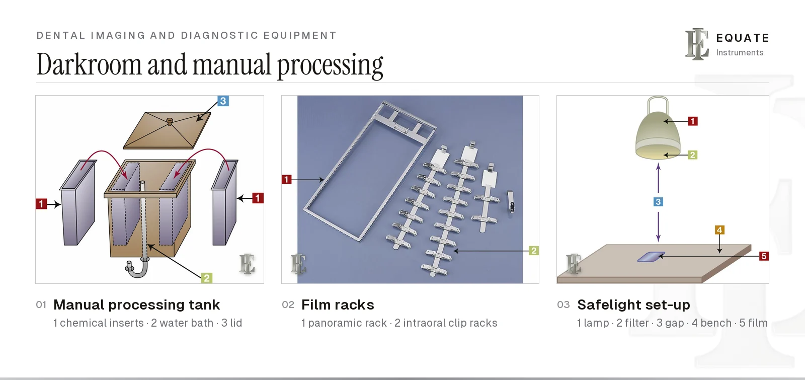
Film is still used in some clinics and teaching settings, and processing it by hand takes a darkroom with a manual tank. The tank holds separate inserts for developer and fixer, sitting in a bath of running water that both rinses the films and keeps the chemicals at a steady temperature. A lid keeps light out. Development time depends on temperature, so a time-and-temperature chart is followed, and fixing always takes longer than developing. Films then need a thorough final wash before drying.
Unwrapped films go onto racks or hangers whose spring clips hold them apart. Moving films between clips part-way through invites scratches and mix-ups, so a single rack carries them from the developer all the way to the drying stage. Racks come in intraoral sizes with a few to a dozen or more clips, and as larger frames for panoramic film. Stainless steel racks resist the chemicals and are cleaned and disinfected between uses.
The darkroom safelight gives just enough filtered light to work by without fogging film. It must use a filter suited to the film type and sit at a safe distance above the bench, commonly given as about 1.2 metres (4 feet). Even a correct safelight is only safe within limits: closeness and handling time both add up, so open packets and load the rack promptly. A simple coin test (laying a coin on unwrapped film under the safelight, then processing it) shows whether the room is safe.
Used fixer contains silver and the lead foil from film packets is a heavy metal, so both are collected for recycling or disposal as national and local environmental regulations require, never poured away or binned with general waste.
Automatic processors, viewers and duplicators
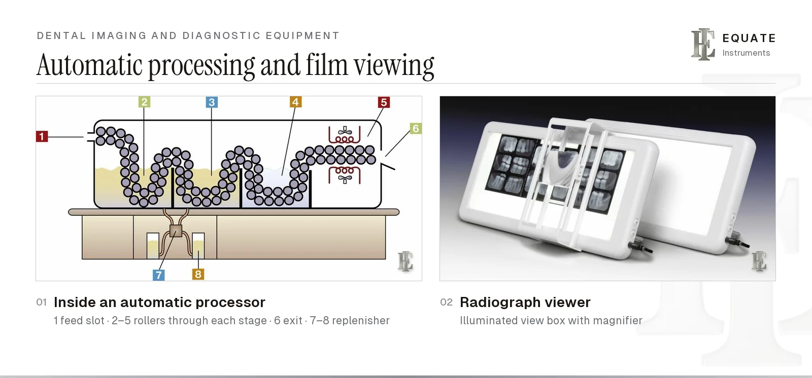
An automatic processor carries each film on rollers through developer, fixer, water and a heated drying section, and delivers it dry at the exit slot, ready to read. Pumps top up the chemicals from replenisher bottles. Processors are used in a darkroom or fitted with a daylight loader, a light-proof box with sleeved hand openings and a filtered viewing window, so films can be unwrapped in a normal room. Use only the chemistry the processor's maker specifies; solutions made up for hand tanks are designed for different temperatures and times. Rollers need regular cleaning, because chemical deposits leave marks on film.
Processed films are read on a view box, an evenly lit panel that is ideally used with surrounding light masked off; some have a built-in magnifier. Copies for referrals or records are made either with double-film packets at the time of exposure or with a film duplicator, which exposes special duplicating film through the original under light. With digital imaging, a copy is simply exported or printed.
Panoramic, cephalometric and cone-beam CT imaging
Extraoral imaging places the receptor outside the mouth. A panoramic unit rotates the tube and receptor around the head to produce one image of both jaws, the teeth, the temporomandibular joints and the sinuses. It is widely used for orthodontic assessment, impacted and third molars, and an overview before surgery. A cephalometric attachment takes a standardised side view of the skull and facial profile, mainly for orthodontic diagnosis and measuring growth. Both can be digital or film-based; combined units do both, and some add cone-beam CT.
Cone-beam CT (CBCT) produces a three-dimensional volume that can be sliced in any plane. It is useful for implant planning, impacted teeth, complex root canal anatomy, some root fractures and pathology. Its dose is generally higher than a panoramic radiograph's, so the European Commission's evidence-based guidelines, Radiation Protection 172 (SEDENTEXCT, 2012), say CBCT should be used only when the clinical question cannot be answered adequately by lower-dose conventional radiography, and then with the smallest field of view and lowest exposure that will do. For scale, FGDP(UK)'s Selection Criteria for Dental Radiography put average UK background radiation at about 2,230 µSv a year. On that yardstick a periapical equals up to about three and a half days of background, a panoramic no more than six days, and a CBCT scan anything from two days to six months, depending on the machine and settings; a chest X-ray is about three days. For related clinical workflows see the orthodontic instruments guide and the oral surgery and extraction instruments guide.
Boyd also covers an enhanced oral assessment aid: a handheld device that shines blue light on the oral mucosa and lets the clinician see the tissue's natural fluorescence through a filter. Areas where fluorescence is lost may need closer investigation. These devices support the conventional visual and tactile examination; they don't diagnose cancer, and a suspicious lesion still needs referral and, where indicated, biopsy. A 2021 Cochrane review by Walsh and colleagues pooled 23 studies of light-based tests in people with visible lesions: they caught most cancers and potentially malignant lesions (sensitivity 0.87) but flagged half of the harmless ones too (specificity 0.50), on low to very low certainty evidence. Its authors concluded that no adjunct test can stand in for a surgical biopsy with histology.
Infection control and care of imaging equipment
CDC guidance treats intraoral radiography accessories as semi-critical items, because they contact mucous membranes: its 2003 dental guidelines say heat-tolerant film-holding and positioning devices should be heat-sterilised between patients, and its current summary puts digital sensors in the same class. In England, HTM 01-05 sets the equivalent decontamination expectations. Follow the manufacturer's instructions and your national rules for each item.
- Heat-tolerant holders (bite blocks, rings, arms, indicator rods): take them apart, clean them, pack them in a pouch or wrap with a process indicator (see the sterilization and protective equipment guide) or load them in an instrument cassette, and steam sterilise them; check the indicators before use.
- Single-use items (bitewing tabs, foam bite blocks, barrier sleeves, film packet wrappers): discard after one patient.
- Digital sensors: cover with a fresh barrier for each patient, then clean and disinfect after removing it, as the manufacturer specifies. The CDC advises heat sterilisation or high-level disinfection where the sensor tolerates it; many don't, so the barrier matters.
- Phosphor plates: seal each plate in a barrier envelope, wipe the envelope before opening it with clean gloves, and never let a contaminated plate go into the scanner. Scratched or creased plates cause artefacts and should be replaced.
- X-ray unit: put barriers on the tube head, PID, control panel and exposure button, or clean and disinfect them between patients.
- Exposed film packets: wipe or remove the outer wrapper so contaminated packets don't enter the darkroom or processor; collect the lead foil separately.
- Mucosal fluorescence devices, and panoramic or CBCT bite blocks, chin rests and head positioners: use a single-use cover or barrier for each patient, then clean and disinfect as the manufacturer specifies.
- Aprons, view boxes and duplicators: clean and disinfect as the manufacturer recommends when they are contaminated.
Choosing imaging accessories: what to check
- Receptor type first: holders, barriers and sizes must suit your film, sensor or phosphor plate system.
- Holder system: paralleling ring-and-arm holders give the most consistent geometry; check that the colour coding matches what your team already uses.
- Sensor fit: holder slots should grip the sensor firmly, leave room for the cable and not press on its edges.
- Reprocessing: choose holders and indicator rods that are labelled for steam sterilisation and come apart for cleaning.
- Collimation: a rectangular PID or a collimator insert cuts patient dose if your unit doesn't have one.
- Barriers: sleeves and envelopes should be the right size for each receptor so they seal fully without stretching.
- Film processing: stainless steel racks or hangers with enough clips for a full series, and fresh chemicals replaced on schedule.
- Compliance: radiation equipment, dosimetry and any shielding have to meet your national radiation protection regulations.
The Equate difference
Equate does not make X-ray units, sensors, film or processors. Its diagnostic range does include 4 stainless steel X-ray film hangers for processing and drying film: a single-hook film clip and racks with 6, 10 and 14 clips (EI-2459 to EI-2462). It also lists 4 endodontic measuring scales for sizing files against a radiograph.
Equate Dental Imaging and Diagnostic Equipment options

X-Ray Film Clip - Single Hook
EI-2459
X-Ray Film Hanger - 6 Clips
EI-2460
X-Ray Film Hanger - 10 Clips
EI-2461
X-Ray Film Hanger - 14 Clips
EI-2462
Endodontic Measuring Scale - Stainless Finger Ring
EI-2918
Endodontic Measuring Ruler - Blue
EI-2919
Endodontic Measuring Ruler - Green
EI-2920
Endodontic Measuring Scale - Pink
EI-2921Browse all diagnostic instruments
Buying in your market
Frequently asked questions
What equipment is needed to take a dental X-ray?
An X-ray unit with a position-indicating device, a receptor (film, a digital sensor or a phosphor plate), a holder or tab to keep the receptor in place, a barrier for digital receptors, and either processing equipment for film or imaging software for digital images.
What do the colours on X-ray film holders mean?
Most paralleling holder systems use blue for anterior periapicals, yellow for posterior periapicals, red for bitewings and green for endodontic views. The colour of the bite block or slot matches the aiming ring so the parts go together correctly.
What is the difference between a digital sensor and a phosphor plate?
A direct digital sensor is a rigid electronic receptor that sends the image to the computer almost immediately. A phosphor storage plate is thin and flexible like film; it holds the image until it is read in a scanner, which then erases it for reuse.
Do patients still need a lead apron for dental X-rays?
The ADA (2024) and the American Academy of Oral and Maxillofacial Radiology (2023) no longer recommend patient aprons or thyroid collars. European guidance (Radiation Protection 136) and the IAEA say routine aprons aren't needed with optimised equipment, but the IAEA still advises a thyroid collar where the thyroid could be in or near the main beam. In the UK, 2020 guidance finds no case for routine patient aprons and limits thyroid shields to views where the gland is unavoidably in the primary beam. National rules decide what a clinic must do, and the IAEA advises offering an apron to any patient who asks.
How often should you have dental X-rays (bitewings)?
It depends on caries risk, not a fixed recall. FGDP(UK) selection criteria suggest bitewings every 6 months for adults and children at high risk and yearly at moderate risk, until no new or active lesions appear. At low risk, adults have them about every 2 years; children about every 12 to 18 months in baby teeth and every 2 years in permanent teeth.
How much radiation is a dental X-ray?
Very little. FGDP(UK) puts a periapical at up to about three and a half days of natural UK background radiation and a panoramic at no more than six days, against about three days for a chest X-ray. A cone-beam CT scan ranges from about two days to six months of background, depending on the machine and settings.
Can you have a dental X-ray when pregnant?
Usually yes. Dental beams are almost never aimed at the abdomen, and FGDP(UK) says there is no need to delay other views, including panoramic and CBCT, until after the birth. UK guidance adds that scatter dose to the foetus is negligible. Because the subject worries many patients, clinicians are advised to offer to postpone radiographs that are not urgent.
Can a light-based oral examination aid tell whether a mouth lesion is cancer?
Not on its own. In people with a visible lesion, a 2021 Cochrane review found light-based tests picked up most cancers and potentially malignant lesions but also flagged about half of harmless ones, on low or very low certainty evidence. They can support a visual and tactile examination, but a suspicious area still needs referral and, where indicated, a biopsy with histology.
How are dental X-ray holders sterilised?
Reusable holders are taken apart, cleaned, packed with a process indicator and steam sterilised between patients. Single-use tabs and foam blocks are discarded. Sensors can't usually go in a steriliser, so they are covered with a fresh barrier for each patient and cleaned and disinfected as the manufacturer specifies.
When is cone-beam CT used in dentistry?
When a three-dimensional view is expected to change treatment, for example implant planning, impacted teeth, complex root canal anatomy or some pathology. European guidelines (Radiation Protection 172) say it should be used only when lower-dose conventional radiography can't answer the question, with the smallest suitable field of view.
References
- Boyd LB. Dental Instruments: A Pocket Guide, 7th edition. Saunders/Elsevier, 2020 (photographs used under licence)
- European Commission. Radiation Protection 136: European guidelines on radiation protection in dental radiology. The safe use of radiographs in dental practice, 2004
- European Commission. Radiation Protection 172: Cone beam CT for dental and maxillofacial radiology. Evidence-based guidelines (SEDENTEXCT), 2012
- International Atomic Energy Agency. Radiation protection of patients in dental radiology
- American Dental Association. ADA releases updated recommendations to enhance radiography safety in dentistry, 2024 (expert panel recommendations published in JADA)
- Centers for Disease Control and Prevention. Sterilization and Disinfection. Dental Infection Prevention and Control
- Centers for Disease Control and Prevention. Guidelines for Infection Control in Dental Health-Care Settings, 2003. MMWR 2003;52(RR-17)
- Department of Health. Health Technical Memorandum 01-05: Decontamination in primary care dental practices, 2013 edition
- Public Health England and FGDP(UK). Guidance Notes for Dental Practitioners on the Safe Use of X-ray Equipment, 2nd edition, 2020
- Faculty of General Dental Practice (UK). Selection Criteria for Dental Radiography, 3rd edition (updated 2018), eds Horner K, Eaton KA
- Walsh T et al. Diagnostic tests for oral cancer and potentially malignant disorders in patients presenting with clinically evident lesions. Cochrane Database of Systematic Reviews 2021, CD010276.pub3
I am a…
Related
- Endodontic Instruments for Root Canal Treatment
- Dental Sterilisation and Protective Equipment
- Orthodontic Instruments: What Each One Does, Stage by Stage
- Oral Surgery and Extraction Instruments
- Basic Dental Instruments: Mirror, Explorer, Cotton Forceps and Handles
- Periodontal Instruments Explained: Probes, Hoes, Knives and Files
- Endodontic Tray Setup: Opening and Sealing a Tooth
- Dental Measuring Ruler
- Basic Dental Tray Setup (Dental Exam Tray)
- Rubber Dam Clamps, Forceps, Punches and Frames: Which to Choose